We are an interdisciplinary group working at the interface of molecular neuroscience, computational biology, and biophysics.
Many of our questions span multiple length and timescales, from ions moving around a single channel to whole tissues, and from microseconds to hundreds of millions of years of evolution. Chemical engineering, a discipline built on connecting molecular processes to the behavior of whole systems, is a remarkably natural home for this work. Although our group is small, it is intellectually diverse, bringing together chemical engineers, physicists, neuroscientists, and computational biologists. Our primary aim is to advance basic understanding, but we also want our work to be useful to others pursuing complementary questions. The group therefore has a highly collaborative ethos. Our research is organized around three areas.
Neurogenomics of development and evolution
The vertebrate brain is built from thousands of neuron types, each with its own molecular identity, shape, and wiring. How does a neuron settle on what it will become? And how has evolution remodeled this cast of cells to suit each species' way of life? We read these histories directly from molecular data, combining single-cell and spatial genomics with statistical inference. Our main model is the retina, the most accessible part of the brain, along with the visual cortex.
We find that neuronal identity is often settled gradually, through an interplay of genetic programs and sensory experience whose balance differs from one cell type to the next (Xie et al., PNAS 2025; Somaiya et al., J. Neurosci. 2025; Yoo et al., bioRxiv 2025). Across vertebrates, many retinal neuron types turn out to be remarkably ancient. Evolution has reshaped vision largely by tuning the abundance, placement, and gene expression of cell types that date back to early vertebrates (Hahn et al., Nature 2023; Tommasini et al., Curr. Biol. 2025).
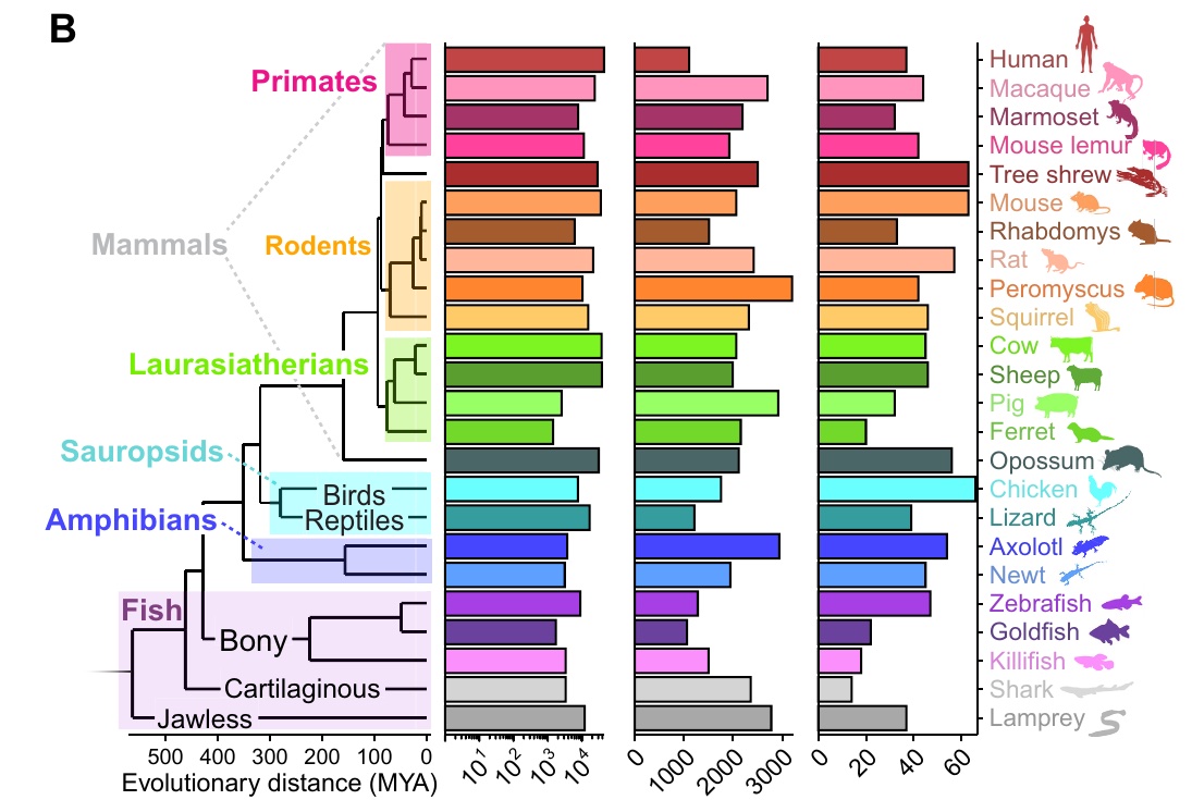
Spotlight. Amacrine cells, the retina's inhibitory interneurons, rival cortical interneurons in their diversity. Led by graduate student Dario Tommasini, we integrated transcriptomic atlases from 24 vertebrate species and identified 42 amacrine types conserved across species. We showed that amacrine diversity scales with that of retinal ganglion cells, and traced the class to an ancient precursor shared with ganglion cells (Tommasini et al., Science Advances 2026).
Key collaborators: Teresa Puthussery (UC Berkeley), Marla Feller (UC Berkeley), Larry Zipursky (UCLA), Nick Tran (Baylor College of Medicine)
Spatial biology of degeneration and aging
In neurodegenerative disease, neurons do not die at random. In glaucoma, the leading cause of irreversible blindness, some retinal ganglion cell types are lost early while others endure. We think part of the answer lies in where a neuron lives, and we build spatial genomic approaches to map cell types across intact tissue and follow their fates in disease.
We are now bringing this spatial view to aging. With Zhigang He's lab, we are asking how the optic nerve changes with age and what limits its ability to regenerate after injury. As part of the Glaucoma Research Foundation's Catalyst for a Cure (CFC4) team, we search for mechanisms that glaucoma shares with other neurodegenerative diseases, including the immune and glial responses that shape neuronal survival (Benhar et al., Nat. Immunol. 2023; Delivanoglou et al., Neuron 2026).
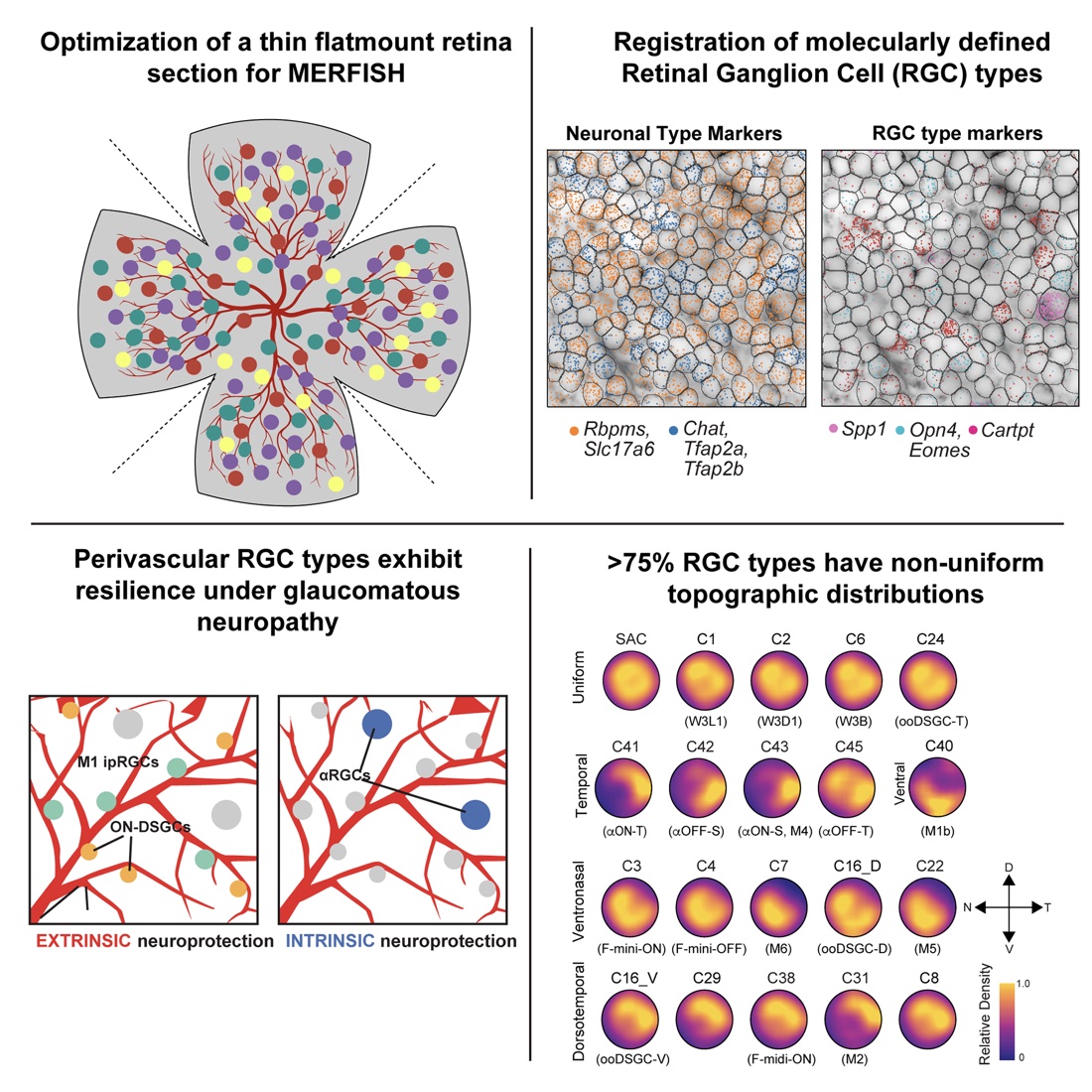
Spotlight. Together with Xin Duan's lab, graduate student Kushal Nimkar adapted MERFISH spatial transcriptomics to thin retinal flatmounts, placing 45 molecularly defined ganglion cell types on a map of the whole retina. More than three-quarters of these types are unevenly distributed. In a glaucoma model, survivors clustered near blood vessels. Perivascular direction-selective and M1 intrinsically photosensitive ganglion cells proved especially resilient, pointing to protection that comes from a neuron's surroundings (Nimkar, Tsai et al., Neuron 2025).
Key collaborators: Xin Duan (UCSF), Zhigang He (Boston Children's Hospital), and CFC4 colleagues Sandro Da Mesquita (Mayo Clinic), Milica Margeta (Mass Eye and Ear), and Humsa Venkatesh (Brigham and Women's Hospital)
Microscopic bioelectricity
Every thought, heartbeat, and sensation begins with ions flowing through channels in a membrane only a few nanometers thick. Yet the textbook picture of neuronal electricity, inherited from Hodgkin and Huxley in the 1950s, treats the membrane as a capacitor and channels as resistors, averaging away the physics around each channel. We start with a simple question: what happens, physically, when a single ion channel opens?
This line of research is a close collaboration with Prof. Kranthi Mandadapu (UC Berkeley). It emerged from our discussions during the COVID-19 pandemic, and the work is carried out jointly by our two groups.
Our theory shows that current through a channel creates a spatially structured, evolving voltage disturbance whose reach is set by the membrane's dielectric properties, geometry, and confinement. It also clarifies when the classical circuit picture emerges (Farhadi et al., Phys. Rev. E 2025; Fernandes et al., Phys. Rev. Research 2026). Electrical and mechanical forces are intertwined: membrane thickness and surface charge govern how electric fields destabilize a bilayer (Ning et al., arXiv 2026).
If neighboring channels feel each other through these fields, the membrane becomes a many-body system. Just as the many forms of ice cannot be deduced from a single water molecule, channel ensembles may support collective bioelectric phases that single-channel models miss. We are now developing a theory of these bioelectric phase transitions and asking how they shape excitability and disease.
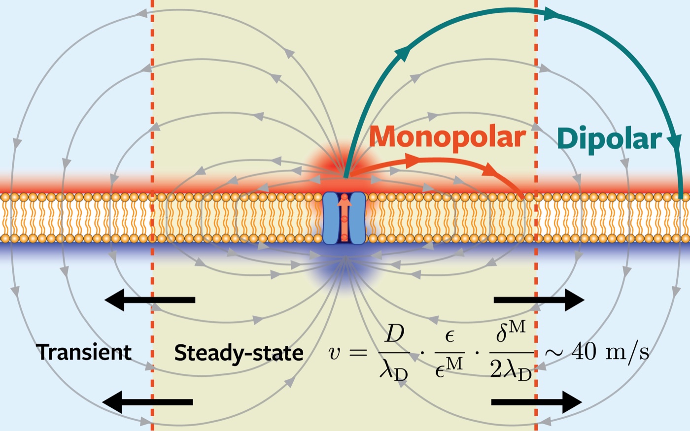
Spotlight. What happens when ions cross a membrane at a single point? Led by postdoc Hyeongjoo Row and graduate student Joshua Fernandes, we used theory and simulation to follow the resulting electric field in space and time. Localized transport generates long-ranged fields with three regimes: a constant-potential near field, an intermediate monopolar region, and a far-field dipolar region. Under sustained transport, the monopolar region expands at roughly 40 m/s in physiological settings, far faster than ions can diffuse (Row, Fernandes et al., Phys. Rev. Research 2025).
Key collaborator: Kranthi Mandadapu (UC Berkeley)
Our primary home is the Department of Chemical and Biomolecular Engineering. We are also affiliated with the Helen Wills Neuroscience Institute, Center for Computational Biology, Berkeley Vision Science, Berkeley Biophysics, QB3, and the Biological Systems and Engineering division at Lawrence Berkeley National Laboratory.